

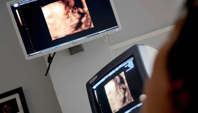
How Ultrasounds Help Monitor Fetal Development

An ultrasound is a standard imaging test during pregnancy, and it creates live pictures of the fetus without using radiation. Since the scan is noninvasive, providers typically repeat it several times over nine months. Each scan gives you and your care team specific information about your baby’s development. This is how ultrasounds help doctors effectively monitor baby development:
Using High-frequency Sound Waves
When a medical provider presses a handheld device called a transducer against your skin, it sends out sound waves. These waves travel at frequencies far above the range of human hearing. The waves bounce off tissues and fluids, and the transducer picks up the returning echoes. Early in pregnancy, the provider may place a slim transducer inside the vagina for a clearer view.
A computer measures the timing of each echo, and it converts the data into an image. Since dense structures like bone appear on an ultrasound as bright white, they reflect more sound. Fluid-filled areas, such as the amniotic sac, show up as black. A standard abdominal scan is typically a short visit.
Visualizing the Fetus
Ultrasound images show the fetus moving in real time. Once the pregnancy reaches about six weeks, your provider can detect a heartbeat. Later scans show much more detail, and the sonographer examines specific body structures one by one. During the mid-pregnancy anatomy scan, the medical provider typically checks these areas:
- Brain and spinal cord
- Heart chambers and major blood vessels
- Kidneys, stomach, and bladder
- Arms, legs, hands, and feet
- Placenta position and umbilical cord
Tracking Fetal Growth
If you are unsure of your conception date, an early ultrasound helps establish your due date. The sonographer measures the length from head to bottom, and this number corresponds closely with gestational age. First-trimester measurements provide the most accurate dating. Your provider uses this date to plan later tests.
Later in pregnancy, providers switch to different measurements, and they record them at each visit. These include head width, head circumference, abdominal circumference, and thigh bone length. A software program combines these numbers so it can estimate the baby’s weight. Your provider may also check the amount of amniotic fluid.
Your provider compares each result against standard growth charts. A single scan offers only a snapshot, but repeated scans reveal a growth pattern over weeks. Estimated weights may differ from actual birth weight. When growth slows or speeds up, your provider may order more frequent scans.
Detecting Complications Early
Routine scans screen for a range of problems. Ultrasounds do not detect every condition, but they flag many concerns early in pregnancy. If a scan shows an unusual finding, your provider may recommend blood work, genetic screening, or a follow-up scan. Scans may reveal conditions such as the following:
- Low or high amniotic fluid levels
- Slowed fetal growth
- Structural differences in the heart or spine
- Potential twins, triplets, or a breech position
Schedule an Ultrasound Near You
Your provider sets the schedule for your ultrasounds. While timing varies, most providers schedule scans during early and mid-pregnancy. Bring your referral and insurance card to your appointment. Call an OBGYN clinic today, and a staff member will review available appointment times with you.